Calcific Tendonitis
What is Calcific Treatment ?
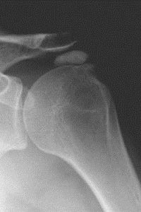
Calcific tendonitis is an important cause of sudden shoulder pain. There may not be any history of trauma. Stage of the disease process, size and location of the calcium deposit determine the presentation. Calcium deposit increase the bulk of the tendon causing subacromial impingement.
Sometimes the calcium crystals leak into the subacromial bursa. This generates chemical inflammation. Such cases present as dull aching pain with episodes of acute pain. It can be confused with frozen shoulder due to reduced movements from pain.
How Is It Diagnosed ?
What Are The Predisposing Factors ?
Will It Resolve Itself ?
What Are The Treatments Options ?
Initially this can be treated with Painkillers and anti-inflammatory medications. Physiotherapy can be useful to keep your shoulder strong and flexible. But it may be difficult in acute phase due to pain inhibition. Steroid injections have a temporary effect to reduce the inflammation and pain.
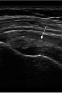
Another procedure called barbotage involves breaking down the deposit under ultrasound guidance. However, this leaves residual calcium crystals in the bursa that generates inflammation and pain.
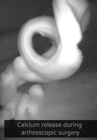
Surgery is required if the pain is not controlled with conservative means. It is performed through keyhole (arthroscopy) and can be done as a day case. You need an experienced shoulder surgeon to carry out this operation. Watch this video to understand about calcium deposits in shoulder in more detail.


The aim of surgery is:
- To reduce the bulk of calcium deposit to prevent subacromial impingement.
- Reduce the pressure effect of the deposit itself (pic 3).
The recovery is good and most patients feel better within 6 weeks.

